
Chinese Journal of Chromatography ›› 2026, Vol. 44 ›› Issue (5): 499-514.DOI: 10.3724/SP.J.1123.2025.10003
• Reviews • Previous Articles Next Articles
YANG Yuanchao1, XIAO Renfang1, WU Yangtao2, BAI Li2, WANG Xingya1, ZHAI Jingbo1,2,3,4,*( )
)
Received:2025-10-06
Online:2026-05-08
Published:2026-05-07
Supported by:CLC Number:
YANG Yuanchao, XIAO Renfang, WU Yangtao, BAI Li, WANG Xingya, ZHAI Jingbo. Polydimethylsiloxane microfluidic aptasensor: opening a new era of biomarker point-of-care testing[J]. Chinese Journal of Chromatography, 2026, 44(5): 499-514.
Add to citation manager EndNote|Ris|BibTeX
URL: https://www.chrom-china.com/EN/10.3724/SP.J.1123.2025.10003
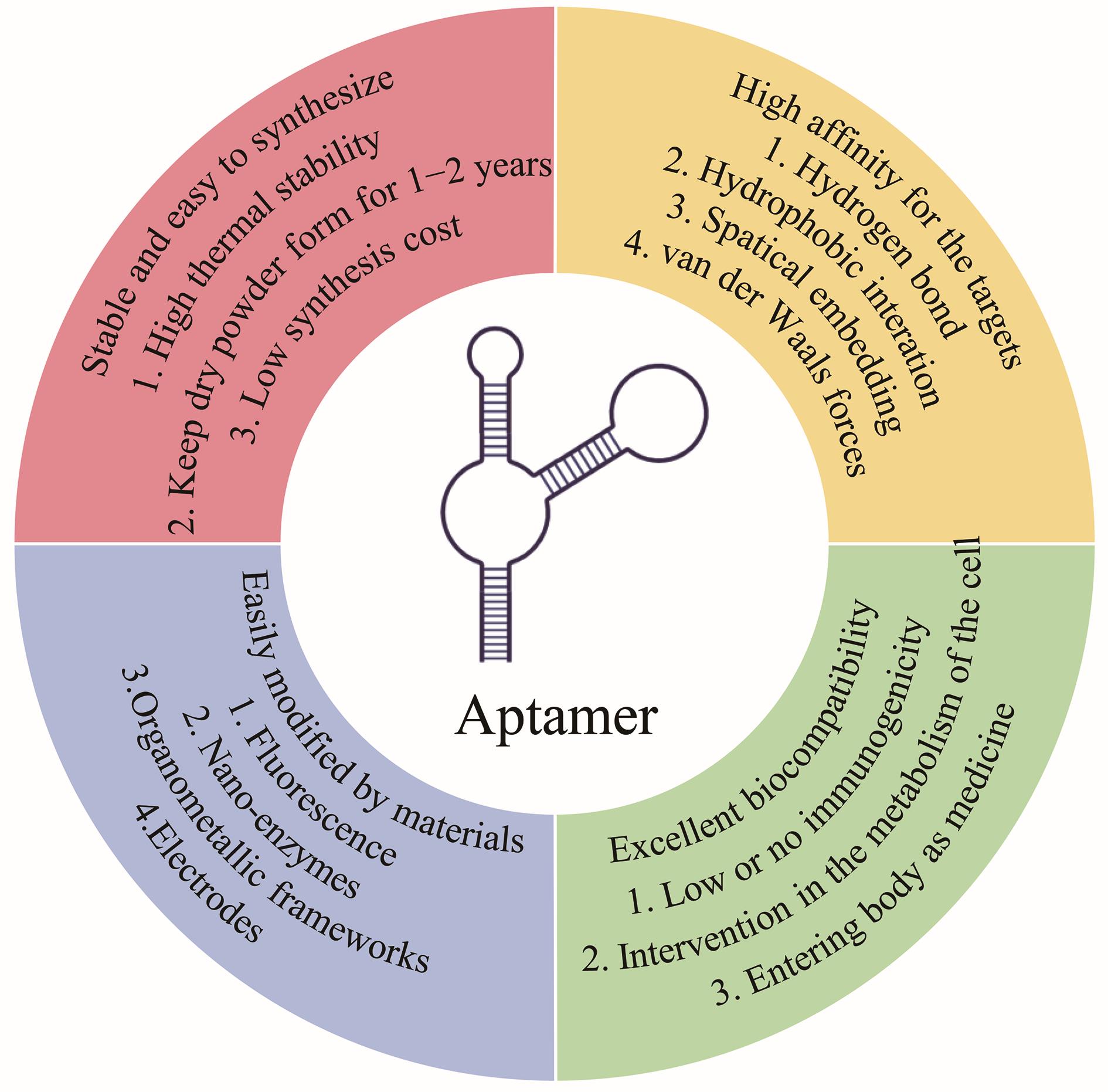
Fig. 1 Aptamer characteristics
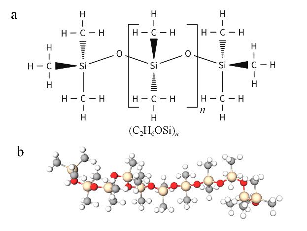
Fig. 2 Structure of polydimethylsiloxane (PDMS)a. chemical structure and chemical formula of PDMS; b. 3D ball-and-stick structure of PDMS.
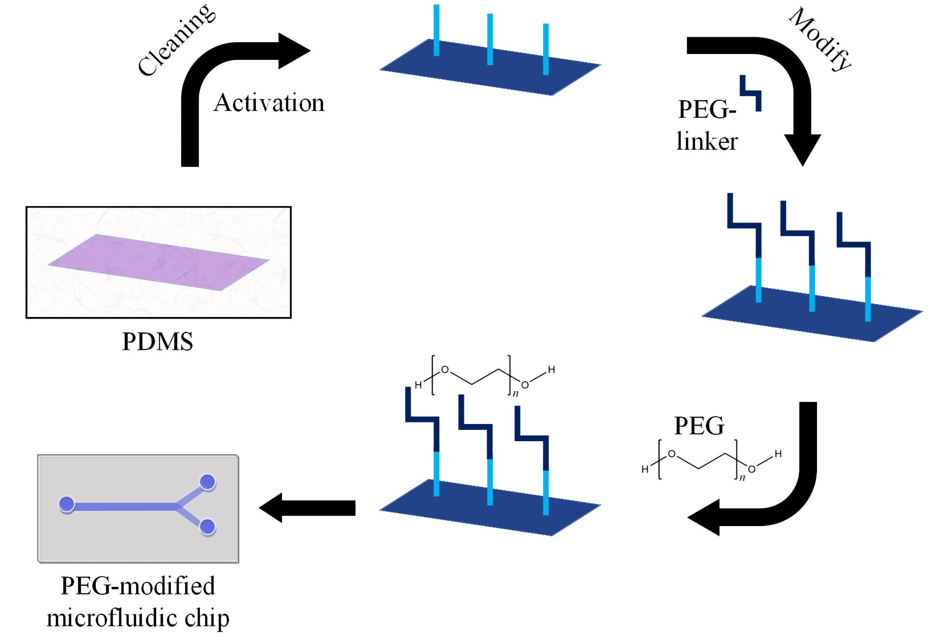
Fig. 3 PEG modified PDMSPEG: polyethylene glycol.
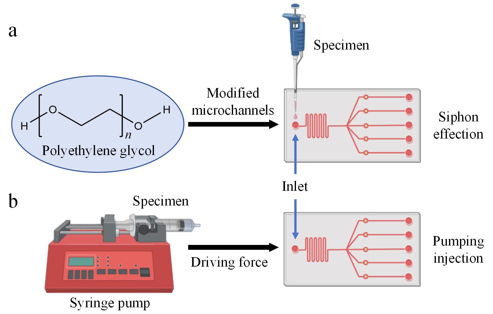
Fig. 4 Differences between hydrophilic and hydrophobic PDMS microfluidic chipsa. The chip modified with PEG hydrophilic can directly use the siphon effect to drive the fluid flow in the channel. b. The chip without hydrophilic modification needs to use the pumping effect to drive the fluid flow in the channel.
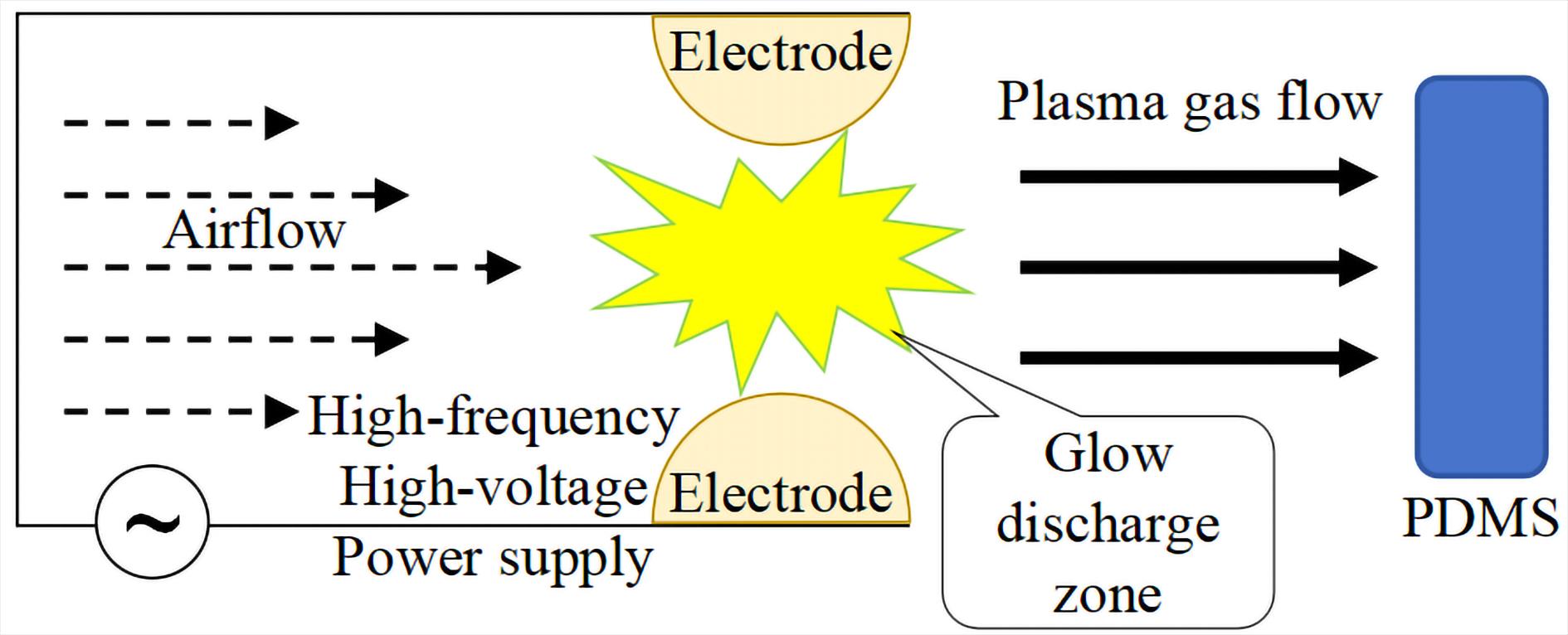
Fig. 5 Schematic diagram of plasma bombardment on PDMS material’s surface
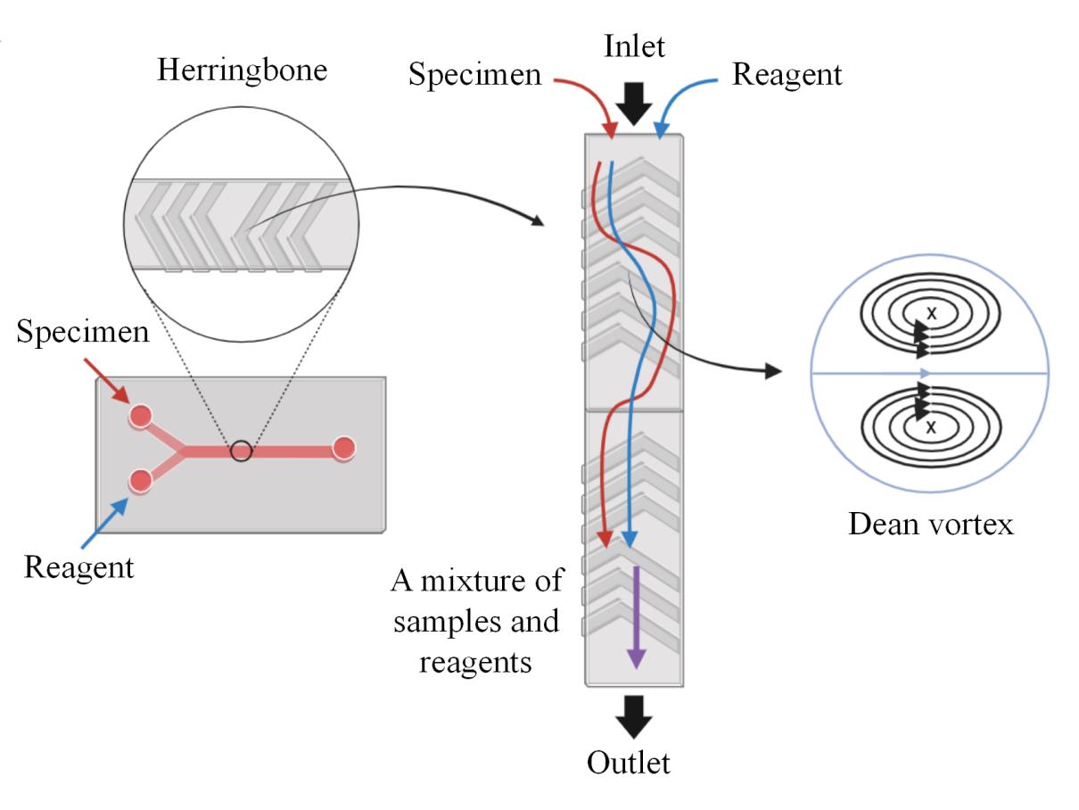
Fig. 6 Schematic diagram of Dean vortex generated by staggered herringbone mixer (SHM) and liquid
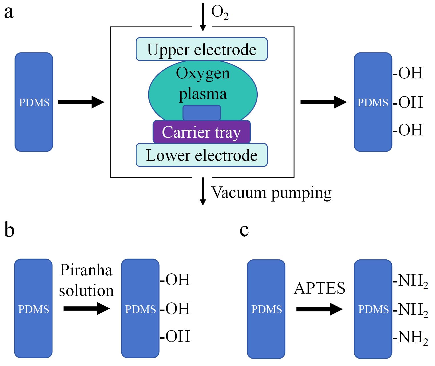
Fig. 7 PDMS surface pretreatment methoda. surface oxygen plasma ablation method; b. piranha solution chemical etching method; c. silanization reagent 3-aminopropyl triethoxy silane (APTES) modification method.
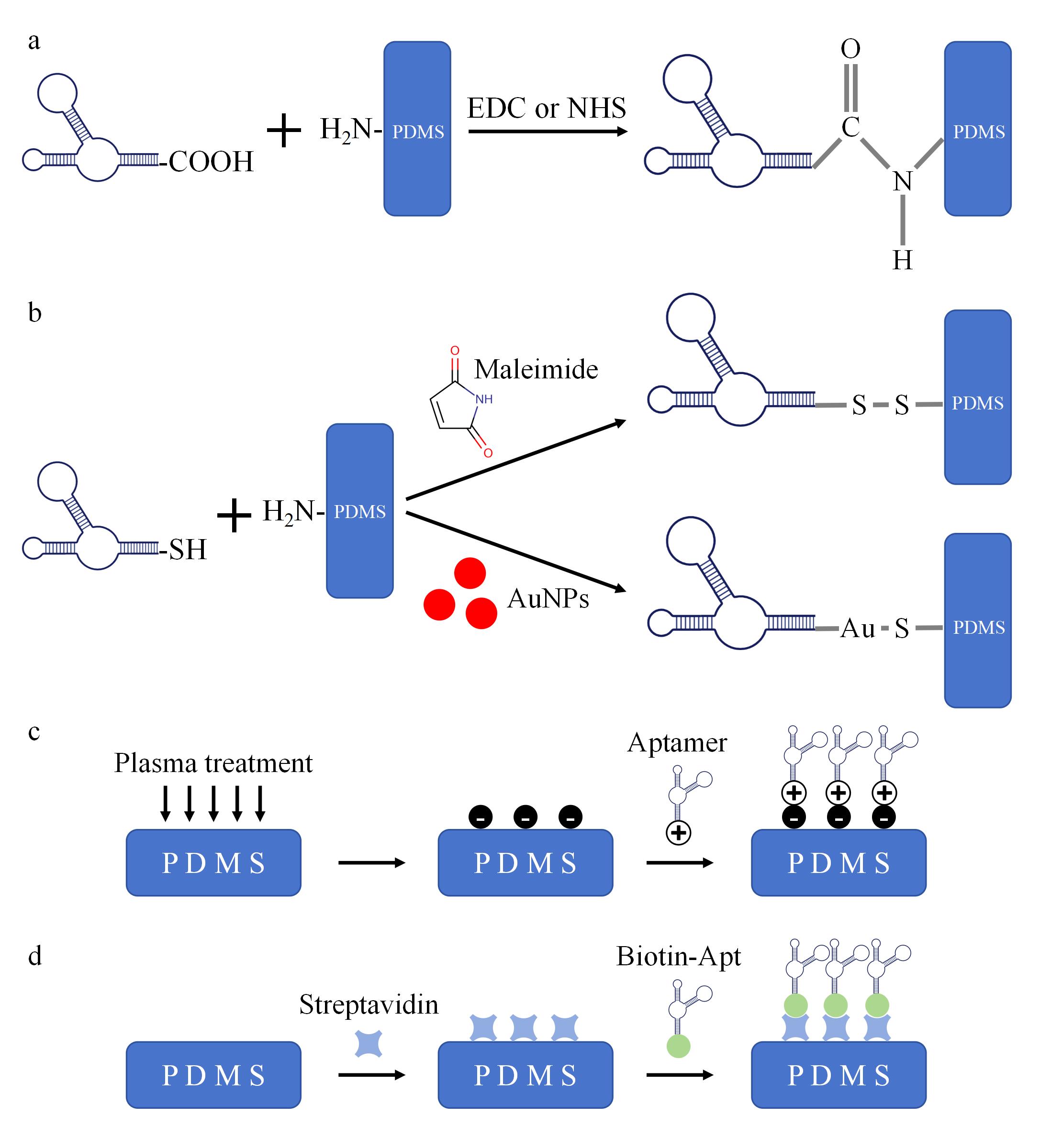
Fig. 8 Schematic diagram of covalent and non-covalent fixationa. arboxyl-modified aptamers are activated via 1-(3-dimethylaminopropyl)-3-ethylcarbodiimide (EDC) or N-hydroxy succinimide (NHS), upon contacting PDMS, their carboxyl groups form amide bonds with amino groups on the PDMS surface, establishing stable attachment. b. Malonamide or AuNPs mediate carboxyl-modified aptamers to bind with amino groups on PDMS surfaces, forming -S-S- bonds or Au-S bonds. c. After plasma treatment, PDMS surfaces acquire negative charges that attract positively charged aptamers through electrostatic adsorption, securing them onto the surface. d. PDMS surfaces are pre-modified with streptavidin (SA), which binds to biotin in biotin-modified aptamer (Biotin-Apt) via specific biotin-helix-ligand interactions, achieving aptamer immobilization.
| Detection method | Strategies of specificity | Targets | Aptamer modifications | Linear range | LOD | Ref. |
|---|---|---|---|---|---|---|
| Colorimetric | visual inspection or quantitative analysis by scanner | thrombin | AgNPs | 20-5000 pmol/L | 20 pmol/L | [ |
| Fluorescence | measured using a cell imaging microplate detection system | NEV released miR | / | 105-109 EVs/mL | miR-223-3p: 0.61 fmol/L; miR-425-5p: 0.24 fmol/L | [ |
| collecting fluorescence signals for quantitative analysis | S.A, S.T, V.P | AuNPs | S.A and S.T: 102-108 CFU/mL;V.P: 101-108 CFU/mL | S.A: 36 CFU/mL; S.T: 39 CFU/mL, V.P: 7 CFU/mL | [ | |
| Chemi-luminescence | biotin-labeled hairpin probes enable HCR amplification and detection | miR-21, miR-155 | / | 1 fmol/L-10 pmol/L | miR-21: 0.39 fmol/L; miR-155: 0.49 fmol/L | [ |
| Raman scattering | rectangular detection zone via magnetic aggregation concentration detection | AFP, MnSOD | 4-MBA, DTNB | 10-12-10-6 g/mL | AFP: 5.89 pg/mL; MnSOD: 6.23 pg/mL | [ |
| SHM promotes hybridization, enabling signal readout via nanoprobes | prostate cancer cell exosomes | AuNPs | 1-105 particles/μL | 1 particle/μL | [ | |
| target binding to the aptamer replaced FAM, resulting in signal attenuation | urea and UA | 5′-FAM | / | / | [ | |
| Electro-chemistry | target binding to the aptamer impedes electron transfer | C. parvum oocysts | / | 10-10000 oocysts/mL | 10 oocysts/mL | [ |
| differential pulse voltammetry | cortisol hormone | / | 1 pmol/L-1 μmol/L | 0.2 pmol/L | [ | |
| Dual-modal | photoelectrochemistry and fluorescence | NSCLC cells NCI-H460, NCI-H1650 | AuNPs | NCI-H460: 50-1×10⁶ cells/mL; NCI-H1650: 50-5×10⁵ cells/mL | NCI-H460: 16 cells/mL; NCI-H1650: 15 cells/mL | [ |
| near-infrared photoelectrochemical and fluorescence imaging with supplementary SHM | liver cancer CTCs cells HepG2 and Hep3B | AuNPs | 100-5×10⁶ cells/mL | HepG2: 8 cells/mL; Hep3B: 6 cells/mL | [ |
Table 1 Performance indexes of biomarker aptasensor based on PDMS microfluidic chip
| Detection method | Strategies of specificity | Targets | Aptamer modifications | Linear range | LOD | Ref. |
|---|---|---|---|---|---|---|
| Colorimetric | visual inspection or quantitative analysis by scanner | thrombin | AgNPs | 20-5000 pmol/L | 20 pmol/L | [ |
| Fluorescence | measured using a cell imaging microplate detection system | NEV released miR | / | 105-109 EVs/mL | miR-223-3p: 0.61 fmol/L; miR-425-5p: 0.24 fmol/L | [ |
| collecting fluorescence signals for quantitative analysis | S.A, S.T, V.P | AuNPs | S.A and S.T: 102-108 CFU/mL;V.P: 101-108 CFU/mL | S.A: 36 CFU/mL; S.T: 39 CFU/mL, V.P: 7 CFU/mL | [ | |
| Chemi-luminescence | biotin-labeled hairpin probes enable HCR amplification and detection | miR-21, miR-155 | / | 1 fmol/L-10 pmol/L | miR-21: 0.39 fmol/L; miR-155: 0.49 fmol/L | [ |
| Raman scattering | rectangular detection zone via magnetic aggregation concentration detection | AFP, MnSOD | 4-MBA, DTNB | 10-12-10-6 g/mL | AFP: 5.89 pg/mL; MnSOD: 6.23 pg/mL | [ |
| SHM promotes hybridization, enabling signal readout via nanoprobes | prostate cancer cell exosomes | AuNPs | 1-105 particles/μL | 1 particle/μL | [ | |
| target binding to the aptamer replaced FAM, resulting in signal attenuation | urea and UA | 5′-FAM | / | / | [ | |
| Electro-chemistry | target binding to the aptamer impedes electron transfer | C. parvum oocysts | / | 10-10000 oocysts/mL | 10 oocysts/mL | [ |
| differential pulse voltammetry | cortisol hormone | / | 1 pmol/L-1 μmol/L | 0.2 pmol/L | [ | |
| Dual-modal | photoelectrochemistry and fluorescence | NSCLC cells NCI-H460, NCI-H1650 | AuNPs | NCI-H460: 50-1×10⁶ cells/mL; NCI-H1650: 50-5×10⁵ cells/mL | NCI-H460: 16 cells/mL; NCI-H1650: 15 cells/mL | [ |
| near-infrared photoelectrochemical and fluorescence imaging with supplementary SHM | liver cancer CTCs cells HepG2 and Hep3B | AuNPs | 100-5×10⁶ cells/mL | HepG2: 8 cells/mL; Hep3B: 6 cells/mL | [ |
|
| [1] | WU Weixiang, DIAO Fuqiang, GUO Junfei, GU Chunming, WU Lihong, LUO Mingyong. Lipid metabolomics-based biomarker analysis of neonatal sepsis in serum and cerebrospinal fluid [J]. Chinese Journal of Chromatography, 2026, 44(3): 286-295. |
| [2] | YAO Zhenyu, WANG Yufei, ZHU Xiaowen, YANG Hong, WU Jia, ZHANG Guojun. Application of chromatography-mass spectrometry in the clinical analysis of multiple sclerosis [J]. Chinese Journal of Chromatography, 2026, 44(3): 226-233. |
| [3] | ZHANG Yufan, XIA Jingwen, ZHU Jiayuan, ZENG Maosheng, BAI Jingyi, XU Qin, GAO Hang. Application of molecularly imprinted polymers-based sensors for determination of acute coronary syndrome biomarkers [J]. Chinese Journal of Chromatography, 2026, 44(2): 180-190. |
| [4] | WANG Mingwei, TIAN Tao, BAI Ligai, HAN Dandan, YAN Hongyuan. Preparation of superhydrophilic imprinted resin-graphene aerogel composite and its precise recognition of tumor biomarker in urine [J]. Chinese Journal of Chromatography, 2026, 44(2): 191-200. |
| [5] | ZHU Ran, CAI Ganping, ZHENG Haijiao, JIA Qiong. Research progress of molecular imprinting technology in the field of disease diagnosis and therapy [J]. Chinese Journal of Chromatography, 2026, 44(1): 2-16. |
| [6] | QIAN Jiankun, HE Runming, FANG Ke, LI Chenlong, BAO Shan, GU Wen, TANG Song. Progress in the metabolic and biotransformation of polycyclic aromatic hydrocarbons and their derivatives in humans [J]. Chinese Journal of Chromatography, 2025, 43(6): 571-584. |
| [7] | ZHANG Weiya, CHEN Pin, XIE Weixin, GAO Xuanbo, ZHANG Wanfeng, DAI Wei, LIN Siyuan, ZHU Shukui. Tracing the origin of crude oil based on fingerprint profiles obtained by comprehensive two-dimensional gas chromatography-time-of-flight mass spectrometry [J]. Chinese Journal of Chromatography, 2025, 43(6): 688-695. |
| [8] | HUANG Yuxia, WANG Haiyan, ZHANG Yihan, LIN Yifei, QIAO Xiaoqiang, HU Lianghai. Bibliometric analysis of exosomes in the biomarker research field [J]. Chinese Journal of Chromatography, 2025, 43(5): 498-507. |
| [9] | WANG Haiyan, XIE Peijuan, QIAO Xiaoqiang, ZHANG Liyuan. Typical strategy and research progress of efficient isolation methods of exosomes based on affinity interaction [J]. Chinese Journal of Chromatography, 2025, 43(5): 413-423. |
| [10] | BU Caiting, ZHU Xuedong, ZHANG Qianying, SHAO Wenya. Recent advances on the role of exosomes in neurodegenerative diseases [J]. Chinese Journal of Chromatography, 2025, 43(5): 487-497. |
| [11] | SU Yating, QIAN Xiaohong, QIN Weijie. Research advances of liposomes and exosomes in drug delivery and biomarker screening [J]. Chinese Journal of Chromatography, 2025, 43(5): 472-486. |
| [12] | FENG Zhuangzhuang, LIN Xiao, BAO Dejun, HU Xiaojian, ZHANG Haijing, ZHU Ying, ZHANG Xu. Determination of four oxidative stress biomarkers in human urine using solid-phase extraction coupled with ultra performance liquid chromatography-tandem mass spectrometry [J]. Chinese Journal of Chromatography, 2025, 43(4): 317-325. |
| [13] | XU Fei, LIU Tianping, GUAN Yajin, HAO Weichao, WEN Dingsheng, XIE Shuilin, BIE Yanan. Analysis of ischemic stroke biomarkers based on non-targeted metabolomics [J]. Chinese Journal of Chromatography, 2025, 43(2): 139-147. |
| [14] | ZHI Mengxue, WANG Jianshe. Advances in the applications of exposomics in the identification of environmental pollutants and their health hazards [J]. Chinese Journal of Chromatography, 2024, 42(2): 142-149. |
| [15] | HAO Qilong, WANG Jing, YU Liqing, ZHANG Haixia. Graphitic carbon nitride/metal-organic framework microextraction fiber for enriching volatile organic compounds in the exhaled breath of patients with lung cancer [J]. Chinese Journal of Chromatography, 2024, 42(12): 1189-1195. |
| Viewed | ||||||
|
Full text |
|
|||||
|
Abstract |
|
|||||


